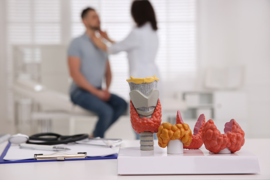
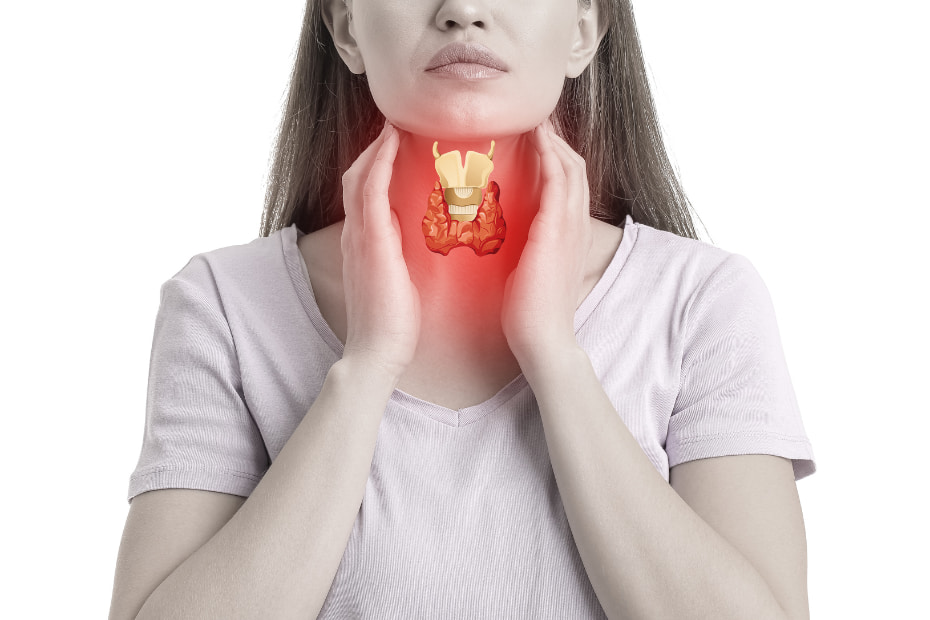
Thyroid tests are essential diagnostic procedures used to evaluate the condition and performance of the thyroid gland. These tests aid in detecting and diagnosing various thyroid disorders, such as hypothyroidism, hyperthyroidism, and thyroid nodules. They encompass a range of methods, including blood tests, imaging techniques, and tissue analysis. Blood tests assess hormone levels and provide insights into overall thyroid function. Imaging tests offer detailed visualizations of the thyroid gland, aiding in identifying structural irregularities. Tissue analysis involves examining thyroid cells to determine if any cancerous or suspicious growths are present. These various thyroid tests are crucial in diagnosing thyroid conditions and guiding appropriate treatment plans.
Blood Tests for Thyroid Function
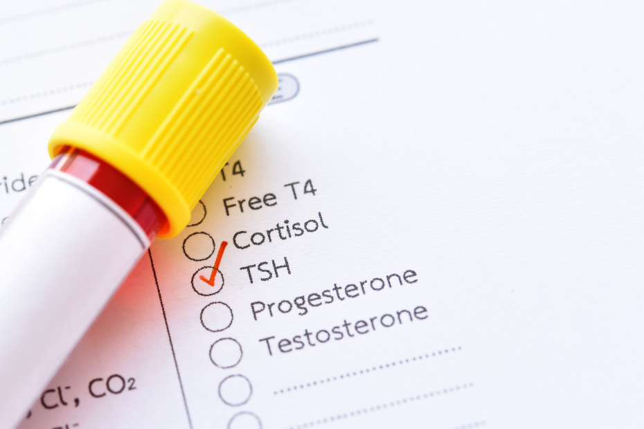
Blood tests for thyroid function are standard diagnostic tools used to evaluate the health and performance of the thyroid gland. These tests measure various thyroid hormones and provide valuable insights into the overall functioning of the thyroid.
TSH (Thyroid Stimulating Hormone) test
The TSH (Thyroid Stimulating Hormone) test is widely used to assess thyroid function by measuring the level of TSH in the blood. The pituitary gland produces TSH and stimulates the thyroid to produce and release thyroid hormones (T3 and T4). Elevated TSH levels indicate an underactive thyroid (hypothyroidism), while low TSH levels suggest an overactive thyroid (hyperthyroidism), as the pituitary gland adjusts hormone production accordingly.
T4 (Thyroxine) and T3 (Triiodothyronine) tests
T4 and T3 tests assess the levels of thyroid hormones in the blood, including both bound and free fractions. T4 serves as the precursor to the more active T3, and these hormones play a vital role in regulating metabolism. Elevated total T3 or T4 levels may indicate hyperthyroidism, while lower levels can indicate hypothyroidism
Free T4 and Free T3 tests
Free T4 and Free T3 tests specifically evaluate the levels of unbound, active thyroid hormones in the blood, providing a more accurate assessment than total T4 and T3 tests. Since they are unaffected by protein fluctuations, these tests are valuable for diagnosing and monitoring thyroid disorders. Changes in free T4 and free T3 levels can indicate early stages of thyroid dysfunction and help assess the effectiveness of treatment.
Reverse T3 (rT3) test
The Reverse T3 (rT3) test measures the levels of an inactive form of T3 that can interfere with free T3’s activity. It helps assess the conversion of T4 to active T3 in the body. Elevated rT3 levels may indicate factors like chronic illness or stress impacting the conversion process. Still, this test is not typically recommended for routine thyroid screening due to its lower reliability than other thyroid function tests.
Thyroid autoantibodies test
These tests detect autoimmune thyroid disorders such as Hashimoto’s thyroiditis and Graves’ disease. Autoimmune thyroid disorders occur when the immune system mistakenly attacks the thyroid gland, leading to either under- or overproduction of thyroid hormones.
There are three types of thyroid autoantibodies tests:
- Thyroid peroxidase (TPO) antibodiesTPO antibodies are found in individuals with Hashimoto’s thyroiditis, the most common cause of hypothyroidism. High levels of TPO antibodies indicate the presence of this autoimmune disorder.
- Thyroglobulin (Tg) antibodiesTg antibodies are measured to assess autoimmune thyroid dysfunction. They can be present in people with Hashimoto’s thyroiditis or Graves’ disease.
- TSH receptor (TSHR) antibodiesTSHR antibodies are associated with Graves’ disease, an immune system disorder that results in hyperthyroidism. The presence of TSHR antibodies can help confirm the diagnosis of Graves’ disease.
Various blood tests can measure thyroid function and detect imbalances or deficiencies of thyroid hormones. If you’re experiencing thyroid disorder symptoms, you must talk to your doctor, who may recommend one or more of these tests to help diagnose and manage your condition.
Imaging Tests for Thyroid Evaluation
Imaging tests are essential for evaluating the thyroid gland, which regulates metabolism through hormone secretion. They aid in diagnosing thyroid disorders, including hyperthyroidism, hypothyroidism, thyroid nodules, and thyroid cancer.
Ultrasound of the thyroid gland
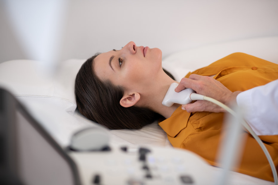
Thyroid ultrasound is a non-invasive imaging test that uses sound waves to visualize the thyroid gland’s size, shape, and structure. It helps identify nodules, cysts, and enlarged lymph nodes, providing valuable information for diagnosing thyroid disorders. The procedure is safe, painless, and typically takes 20 to 30 minutes, involving the application of gel on the skin and using a transducer to create images on a monitor.
Thyroid scintigraphy (nuclear medicine scan)
Thyroid scintigraphy, a nuclear medicine scan, uses a small amount of radioactive material and a specialized camera to assess thyroid gland function. It is instrumental in diagnosing hyperthyroidism and distinguishing between benign and malignant nodules. By visualizing the absorption and activity of the radioactive material, thyroid scintigraphy provides valuable information about the functionality of the thyroid gland. Although generally safe, the test is not recommended for pregnant or nursing women due to potential risks to the fetus or nursing child.
CT (computed tomography) scan
A CT (computed tomography) scan uses X-rays to create detailed cross-sectional images of the thyroid gland and adjacent structures. Although not as common as other thyroid imaging methods, a CT scan can provide valuable information about thyroid abnormalities, including cancer, tumor size, surrounding tissue invasion, and enlarged lymph nodes. It’s important to note that CT scans involve ionizing radiation, which carries a potential risk of developing cancer from repeated exposure. During the scan, the patient lies on a table that slides through the CT scanner while X-ray images are taken and compiled into a comprehensive picture of the thyroid and surrounding areas.
MRI (magnetic resonance imaging) of the thyroid
MRI (magnetic resonance imaging) is a safe imaging technique that uses powerful magnets and radio waves to create detailed images of the thyroid gland without ionizing radiation. It provides valuable information about size, structure, blood flow, and thyroid abnormalities, such as nodules, cysts, and tumors. However, patients with certain metallic implants or devices should avoid MRI scans due to the strong magnets used. During the procedure, the patient lies on a table that slides into a tube-shaped MRI scanner, and a contrast material called gadolinium may be administered to enhance thyroid visibility.
PET (positron emission tomography) scan
PET (positron emission tomography) scans are nuclear medicine imaging tests that assess the metabolic activity of tissues using a radioactive substance. In thyroid evaluation, they are commonly used for diagnosing and staging thyroid cancer, detecting cancer spread to lymph nodes or distant organs, and determining the metabolic activity of nodules to assess their malignancy. A radioactive substance called a radiotracer is injected during a PET scan, and the PET scanner creates detailed images of metabolic activity. These scans involve radiation exposure and are not recommended for pregnant or nursing women. They are typically performed when other imaging methods are insufficient or in suspected thyroid cancer recurrence cases.
Fine Needle Aspiration Biopsy (FNAB) and other Biopsy Techniques
A biopsy is a diagnostic procedure in which a small amount of tissue or cells is removed from the body to be examined under a microscope to diagnose a disease, especially cancer. Fine Needle Aspiration Biopsy (FNAB) is one such technique, and there are others, such as Core Needle Biopsy (CNB) and Open Surgical Biopsy, each of which differs in the way samples are obtained and the type of needles used.
Fine Needle Aspiration Biopsy (FNAB) is a minimally invasive procedure that involves extracting a small tissue or fluid sample from a palpable mass for microscopic examination. It is commonly used for diagnosing lumps or nodules in the breast, thyroid, lymph nodes, and other soft tissue areas. FNAB provides rapid, economical, and safe results, though it may have limitations in cases where the sample size is insufficient or inconclusive. The procedure is typically performed under local anesthesia and causes minimal pain due to the thin needle used.
Core needle biopsy (CNB)
Core Needle Biopsy (CNB), also called cutting needle biopsy, is a minimally invasive procedure that uses a larger hollow needle with a cutting mechanism to obtain a cylindrical tissue core for examination. CNB provides larger tissue samples than FNAB, allowing for more detailed evaluation and accurate diagnosis. It is commonly used to investigate suspicious breast masses. Still, it can also be performed in other areas like the thyroid, lung, liver, kidney, prostate, or bone, often guided by imaging techniques. While CNB offers the advantages of greater sample size and minimal invasiveness, it may cause more discomfort and bruising than FNAB due to the larger needle size.
Open surgical biopsy
Open Surgical Biopsy, performed under anesthesia, involves making an incision to remove a tissue sample or entire abnormal mass, providing a comprehensive specimen for accurate diagnosis. It is typically used when FNAB or CNB are unsuitable or inconclusive, especially for lesions in challenging locations, non-palpable masses, or cases requiring extensive histopathological assessment. While open surgical biopsies yield larger samples for conclusive results, they carry risks of complications and require longer recovery time and higher costs than less invasive techniques.
Functional Tests for Thyroid Disorders
Thyroid disorders are a common clinical problem affecting millions of people worldwide. These disorders can significantly impact the quality of life, and appropriate diagnosis and treatment are crucial for the effective management of these conditions. This article discusses functional tests that help diagnose and monitor thyroid disorders, including TRH stimulation, thyroid suppression, and iodine uptake tests.
TRH (thyrotropin-releasing hormone) stimulation test
The TRH stimulation test evaluates the pituitary gland’s response to thyroid disorders by administering synthetic TRH and measuring TSH levels in the blood. It helps diagnose secondary hypothyroidism and differentiates it from nonthyroidal disorders affecting TSH levels. However, the test should be avoided in suspected hyperthyroidism or thyroid storm cases to prevent symptom exacerbation.
Thyroid suppression test
The thyroid suppression test evaluates the response of the thyroid gland to the T4 hormone by administering synthetic T4 and measuring TSH, T3, and T4 levels. It helps determine the cause of thyroid dysfunction in patients with goiter or suspected hyperthyroidism. If the thyroid gland continues to produce hormones despite TSH suppression, it suggests an overactive thyroid, while normal suppression indicates proper thyroid function. The test helps diagnose the underlying cause of hyperthyroidism and assess treatment effectiveness.
Iodine uptake test
The iodine uptake test evaluates the thyroid gland’s ability to concentrate iodine by administering radioactive iodine and measuring its uptake using imaging techniques. Increased uptake indicates an overactive thyroid, while decreased uptake suggests hypothyroidism or a non-functioning gland. The test aids in diagnosing thyroid disorders, distinguishing between conditions like Graves’ disease and toxic multinodular goiter, and assessing treatment effectiveness.
Understanding Thyroid Test Results
Thyroid tests are essential diagnostic tools that help healthcare providers understand the functioning of the thyroid gland. The thyroid gland is a butterfly-shaped organ located in the front of the neck that produces hormones that regulate many of the body’s essential functions, including metabolism, body temperature, mood, and energy levels. Understanding thyroid test results is essential for patients with suspicions of a thyroid disorder.
Normal thyroid function ranges
Several different tests can evaluate the function of the thyroid gland, but the most common ones are TSH (Thyroid-Stimulating Hormone), T4 (Thyroxine), and T3 (Triiodothyronine).
Here are the typical normal ranges for each test:
TSH: The normal range for TSH levels is usually between 0.5 to 5.0 mIU/L, though this may vary depending on the laboratory and the specific test used. TSH is the hormone that signals the thyroid gland to produce T3 and T4 hormones.
T4 (Total or Free T4): Total T4 measures all thyroxine in the blood, while free T4 measures only the portion available in the bloodstream for use by the body. Normal levels of T4 in the blood range from 5-12 micrograms per deciliter (mcg/dl). Free T4, which represents the unbound and active form of T4, typically falls within the normal range of 0.8-1.8 nanograms per deciliter (ng/dl) of blood.
T3 (Total or Free T3): Similar to T4, Total T3 measures all T3 hormones circulating in the blood, and Free T3 measures only the portion available in the bloodstream. Normal ranges for Total T3 usually range between 80-220 ng/dL.
Interpreting abnormal test results
Abnormal thyroid test results can indicate various thyroid disorders, including hypothyroidism (underactive thyroid), hyperthyroidism (overactive thyroid), or thyroid cancer. The following information can help interpret abnormal test results:
Elevated TSH levels: An abnormally high TSH level, usually above 4.0 mU/L, indicates that the thyroid gland is not producing enough T3 and T4 hormones, causing hypothyroidism. This could be due to a condition such as Hashimoto’s thyroiditis, an autoimmune disorder that damages the thyroid gland.
Suppressed TSH levels: Low TSH levels (below 0.4 mU/L) indicate the thyroid gland overproduces T3 and T4 hormones, resulting in hyperthyroidism. This could be caused by Graves’ disease or a thyroid nodule that produces excess hormones.
Abnormal T4 and T3 levels: T3 and T4 levels outside the normal ranges may indicate a thyroid disorder, depending on the TSH levels. If TSH levels are normal, it may indicate a pituitary gland dysfunction.
In conclusion, the various diagnostic tests discussed, such as Free T4 and Free T3 tests, Reverse T3 tests, imaging tests (ultrasound, scintigraphy, CT scan, MRI, PET scan), fine needle aspiration biopsy (FNAB), core needle biopsy (CNB), and open surgical biopsy, play crucial roles in the evaluation and diagnosis of thyroid disorders.
These tests provide valuable information about thyroid hormone levels, thyroid structure, function, and abnormalities such as nodules or tumors. Each test has advantages, limitations, and indications, allowing healthcare professionals to tailor the diagnostic approach based on the patient’s needs. Furthermore, these diagnostic procedures aid in determining appropriate treatment strategies, monitoring treatment effectiveness, and identifying potential complications. By utilizing these diagnostic tools effectively, healthcare providers can make accurate diagnoses and provide optimal care for patients with thyroid disorders.
